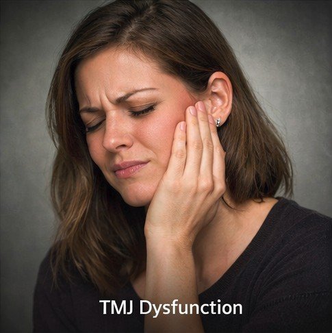
The temporomandibular joint connects the lower jaw to the temporal bone of the skull and is among the most mechanically complex joints in the human body. Every act of speaking, eating, swallowing, or yawning demands precise coordination between these joints and the muscles that drive them. When this coordination is disrupted — whether by joint degeneration, disc displacement, malocclusion, or chronic muscular tension from bruxism — the consequences reach far beyond the jaw itself. Temporomandibular joint disorder, commonly abbreviated TMD, is a significant and frequently underrecognized source of chronic headache, and many patients spend years in diagnostic uncertainty before the jaw origin of their pain is identified.
Pain arising from the TMJ and its surrounding musculature is referred into the temples, the sides of the head, and even the periorbital region through shared trigeminal nerve pathways, creating headaches that closely resemble primary headache disorders in both location and quality. The result is a condition that is routinely misclassified as migraine or tension-type headache, leading to treatments that provide only partial relief because they address the pain perception without targeting the structural source.
Anatomy and Pain Referral Mechanisms
The temporomandibular joint is innervated primarily by the auriculotemporal branch of the trigeminal nerve — the same cranial nerve that mediates sensation across the forehead, temples, cheeks, teeth, and sinuses. This shared neural territory is the anatomical foundation for referred pain from the jaw into the head. The masseter muscle, responsible for biting force, develops myofascial trigger points under the chronic load of clenching or grinding, and these trigger points refer pain directly to the temple and cheekbone. The temporalis muscle fans across the temporal fossa and generates referred pain indistinguishable from tension-type headache when chronically overloaded.
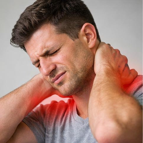
The lateral pterygoid refers pain to the preauricular region and temple when it is in spasm, while the medial pterygoid refers pain behind the ear and to the angle of the jaw. Collectively, the masticatory muscles create a referred pain map that encompasses much of the cranial territory associated with common primary headache disorders. Central sensitization — progressive amplification of pain signals within the central nervous system caused by sustained nociceptive input — adds further complexity in long-standing cases, maintaining headache even when jaw-specific pain is relatively quiescent.
Risk Factors and Clinical Features
Bruxism — the habit of clenching or grinding the teeth, most commonly during sleep — is among the most powerful drivers of TMD-related headache, subjecting the joint and its muscles to repetitive compressive forces that exceed anything generated during normal mastication. Dental malocclusion, direct jaw trauma, inflammatory arthritis affecting the joint, and psychological stress all contribute to TMD through distinct but overlapping mechanisms. Stress deserves particular emphasis, as it drives masticatory muscle hypertonicity through central nervous system mechanisms that operate largely outside conscious awareness.
The clinical features most reliably pointing toward TMD include temporal or preauricular pain that worsens with jaw use, morning headache that peaks upon awakening and correlates with nocturnal bruxism, audible or palpable clicking and popping in the joint, restricted or deviated jaw opening, and tenderness of the masticatory muscles on clinical palpation. Ear symptoms including fullness, low-grade tinnitus, and otalgia without otological pathology are common associated complaints that further support the TMD diagnosis.
Diagnosis
Diagnosis of TMD is primarily clinical. The Diagnostic Criteria for Temporomandibular Disorders provides a validated international framework that categorizes TMD into articular disorders affecting the joint itself and muscular disorders affecting the masticatory muscles. Imaging with MRI provides the most detailed information about disc position and joint integrity, while cone-beam CT superior visualizes bony structural changes. Imaging findings must always be interpreted in clinical context, as asymptomatic disc displacement and degenerative joint changes are prevalent in the general population.
Management of TMD-associated headache is multimodal. Occlusal stabilization splints — custom-made appliances worn over the teeth, typically at night — reduce compressive forces on the joint and provide a physical barrier against bruxism-related damage. Physical therapy targeting both jaw musculature and the frequently involved cervical spine includes myofascial release, trigger point treatment, therapeutic exercise, and postural correction. Botulinum toxin injections into the masseter and temporalis muscles produce clinically meaningful reductions in muscle hypertonicity and associated headache, with effects lasting three to four months per treatment cycle.
Pharmacological support during flares may include NSAIDs, muscle relaxants, or tricyclic antidepressants at low doses. For severe breakthrough pain episodes unresponsive to first-line agents, a physician may recommend combination analgesics. Patients who are directed to purchase fioricet with medical prescription for acute TMD-related headache relief should understand that butalbital-containing medications carry strict frequency limits — typically no more than two days per week — to avoid transforming episodic flare management into medication overuse headache. Those who prefer the convenience of obtaining their prescription remotely can order fioricet online with rx through properly licensed telehealth services that require valid prescriptions and clinical evaluation before dispensing.
Psychological interventions addressing the stress-bruxism-pain cycle are critical for sustained improvement. Cognitive behavioral therapy for pain management, biofeedback training for masticatory muscle relaxation, and stress reduction techniques all have supporting evidence and are particularly valuable for patients in whom psychological factors are prominent. For patients managing periodic flares, a clear pre-agreed acute plan — including criteria for when to buy fioricet at the pharmacy versus when to seek urgent evaluation — reduces the anxiety and decision-making burden that severe pain episodes impose.
Long-Term Prognosis
With comprehensive treatment consistently applied, the prognosis for TMD-associated headache is favorable. Most patients achieve significant reduction in headache frequency and severity with conservative management, and the condition does not typically progress when structural stresses are reduced through splint therapy, ergonomic modification, and appropriate exercises. Long-term success depends on maintaining the behavioral changes — including consistent splint use, regular exercise, and stress management — that address the root drivers of the condition rather than simply suppressing its symptomatic expression.
Sleep hygiene deserves attention in all TMD patients, as sleep quality profoundly influences both nocturnal bruxism intensity and the central pain modulation systems that determine daytime headache burden. Regular aerobic exercise reduces the adrenergic arousal that fuels masticatory muscle hypertonicity and improves the quality of sleep that is essential for tissue recovery. The integration of dental, physical, behavioral, and medical care within a coordinated team approach provides the most reliable foundation for lasting relief.
Temporomandibular joint disorder-related headache and facial pain is a condition of extraordinary clinical complexity, arising from the dysfunction of one of the most mechanically demanding […]
The TMJ-Headache Relationship The temporomandibular joint — universally referred to by its initials TMJ — is one of the most complex and heavily used joints […]
The Paradox at the Heart of Headache Pharmacotherapy One of the most clinically frustrating phenomena in headache medicine is the concept of medication overuse headache […]